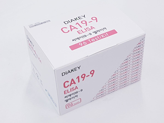
DIAKEY CA19-9 ELISA

| KFDA Registration No : | 14-3154 |
| CAT No : | DEC03 |
| TEST METHOD : | ELISA |
| SAMPLE VOLUME : | 50 ul |
| INCUBATION TIME : | 30'+30'+20'RT |
| STD RANGE : | 0-240 U/ml |
INTENDED USE : Enzyme immunometric assay for quantitative determination of carbohydrate antigen 19-9(CA19-9) in human serum or plasma
INTRODUCTION
Macromolecular tumor markers can be classified into the following major classes: enzymes, hormones and growth factors and their receptors, oncogenic products, viral antigens, oncofetal and surface antigens. With a few exceptions (certain enzymes), most macromolecular tumor markers are glycol-conjugates and a wealth of information is available on these antigens. Therefore, investigations into the expressions of unusual glycoprotein antigens ('markers') associated with tumors and their metastases dominate much of clinical and experimental oncology today.
CA19-9 antigen is a kind of carbohydrate antigen, one of important tumor markers. The monoclonal antibody was obtained by Koprowski et al, in 1979 from hybrids of human colon cancer cell line cultured in vitro. The determinant of CA19-9 was proved to be a sialylated lacto-N-fucopentaose Ⅱ. Its biochemical properties are associated with lewis blood group antigens. Analyses of serum samples by several groups have indicated elevated levels of CA19-9 antigen in several types of malignancies, particularly those of pancreas, stomach and colon, but not in sera of normal individuals. CA19-9 antigen was originally identified as a ganglioside isolated from the SW1116 human colorectal carcinoma cell line, while the serum antigen was characterized as a mucin. It can also be detected on mucin in patients' sera. Sources include normal pancreas, bile duct, and gastric, colic, endometric and salivary epithelia. In healthy individuals, the CA19-9 antigen circulates at low levels, normally less than 37U/mL. Elevated levels have been seen in benign inflammatory diseases of the hepatobiliary tract. Studies have also shown that mucin bearing this antigen was more frequently detected in the sera of patients with pancreatic cancer than any other gastrointestinal carcinoma, including colorectal. It is also found in bile duct, ovarian mucinous cystadenocarcinomas and uterine adenocarcinomas.
PRINCIPLE OF THE ASSAY
The assay is a solid-phase, non-competitive immunoassay (ELISA) based upon the direct sandwich technique. Standards, controls and samples are incubated together with a biotinylated anti-CA19-9 monoclonal antibody in streptavidin coated microplates. CA19-9 present in standards or samples is adsorbed to the streptavidin coated micro strips by the biotinylated anti-CA19-9 Mab during incubation. The strips are the washed and incubated with HRPO labeled anti-CA19-9 Mab. After washing, Chromogen reagent is added to each well and the enzyme reaction is allowed proceed. During the enzyme reaction blue color will develop if antigen is present. The intensity of the color is proportional to the amount of CA19-9 present in the samples. The color intensity is determined in a microplate spectrophotometer at 450nm (reference.620nm).
 HANDLING PRECAUTION
- Do not use mixed reagents from different lots.
- Do not use reagents beyond the expiration date.
- Use distilled water stored in clean container.
- Use an individual disposable tip for each sample and reagent, to prevent the possible cross-contamination among the samples.
- Rapidly dispense reagents during the assay, not to let wells dry out.
 USE PRECAUTION
- Wear disposable globes while handling the kit reagents and wash hands thoroughly afterwards.
- Do not pipette by mouth.
- Do not smoke, eat or drink in areas where specimens or kit reagents are handle.
- Handle samples, reagents and loboratory equipments used for assy with extreme care, as they may potentially contain infectious agents.
- When samples or reagents happen to be spilt, wash carefully with a 1% sodium hypochlorite solution.
- Dispose of this cleaning liquid and also such used washing cloth or tissue paper with care, as they may also contain infectious agents.
- Avoid microbial contamination when the reagent vial be eventually opend or the contents be handled.
- Use only for IN VITRO.